Imaging Case Reports

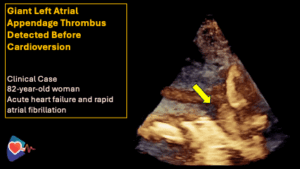
Giant Left Atrial Appendage Thrombus Detected Before Cardioversion
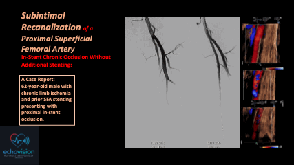
Subintimal Recanalization of a Proximal Superficial Femoral Artery In-Stent Chronic Occlusion Without Additional Stenting
- Subintimal recanalization is a valuable option in chronic SFA in-stent occlusion
- Avoidance of additional stent implantation may reduce metal burden
- Duplex ultrasound is essential for functional assessment
- Good short-term patency can be achieved even with persistent proximal stent thrombosis
This case illustrates the feasibility and effectiveness of a subintimal strategy in complex in-stent chronic occlusions of the SFA.
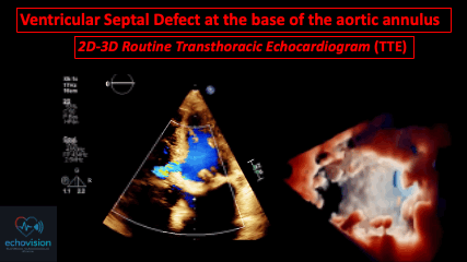
Ventricular Septal Defect at the base of the aortic annulus: 2D-3D Routine Transthoracic Echocardiogram (TTE)
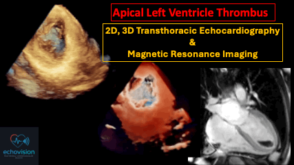
Apical Left Ventricle Thrombus: 2D, 3D Transthoracic Echocardiography & Magnetic Resonance Imaging
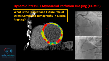
Dynamic Stress CT Myocardial Perfusion Imaging (CT-MPI)
What is the Present and Future role of Stress Computed Tomography in Clinical Practice?
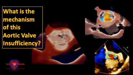
What is the mechanism of this Aortic Valve Insufficiency?
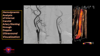
Hemodynamic Analysis of Internal Carotid Artery Stenting through Doppler Ultrasound Visualization
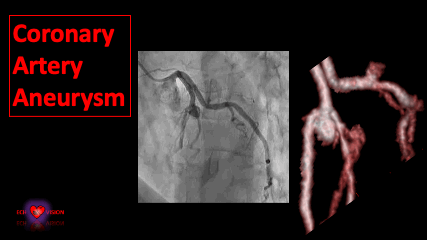
Coronary Artery Aneurysm
A 67-year-old woman, dyslipidemia, with atypical chest pain and slight EKG modification. Coronary artery angiography: Tight stenosis of proximal LAD and D1 with an Aneurysm of 10 mm in diameter overhanging the Bifurcation
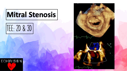
Mitral stenosis in a 70-year-old woman
Mitral stenosis in a 70-year-old woman with pulmonary hypertension: Restrictive movements, particularly of the anterior leaflet. Note the presence of moderate central mitral regurgitation.
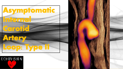
Asymptomatic Internal Carotid Artery Loop: Type II
Boucle Carotide Interne Asymptomatique: Type II
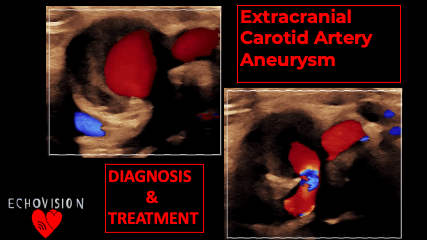
Extracranial Carotid Artery Aneurysm: Diagnosis & Treatment
Anévrysme Carotide Interne : Du Diagnostique au Traitement

MITRAL VALVE ENDOCARDITIS: Very Mobile Posterior Mitral Valve Leaflet Vegetation
Endocardite Mitrale: Végétation très mobile de la valve postérieure